
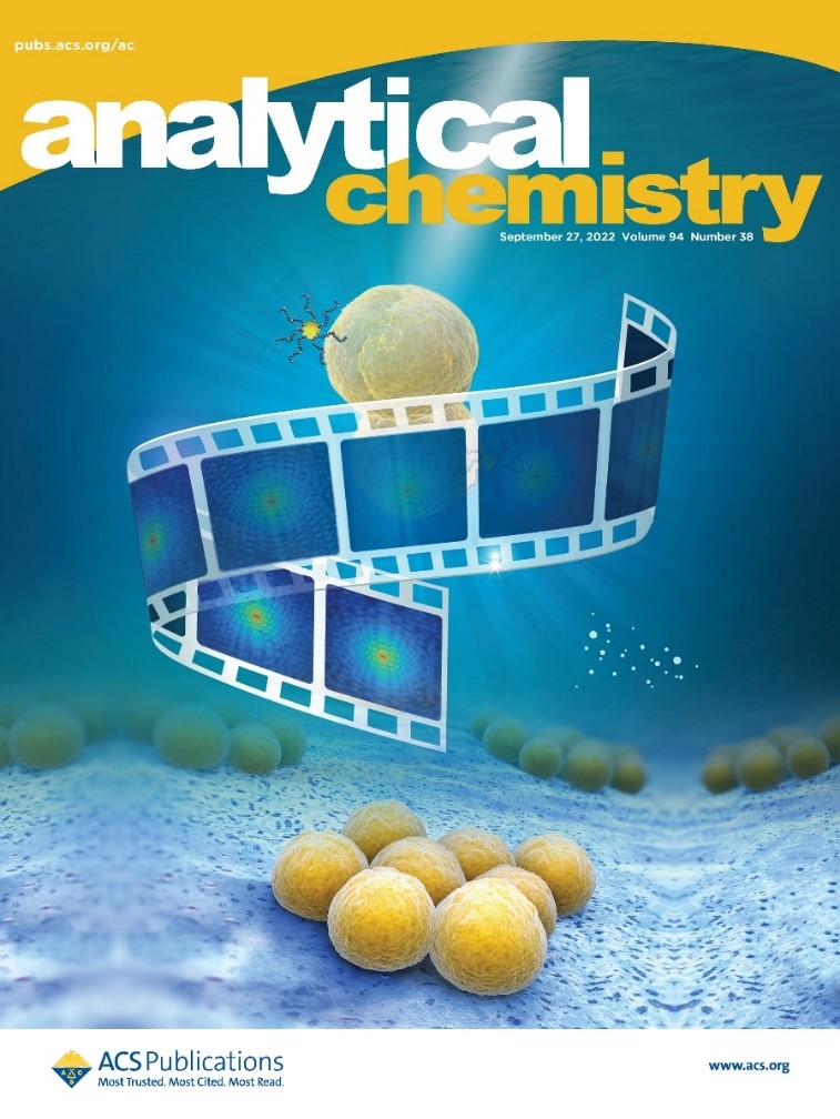
A research team from ShanghaiTech University and Beijing University of Technology achieved a breakthrough in the field of quantitative 3D spatial structural analysis of Staphylococcus aureus to explore the microbe–inhibitor drug interactions at nanoscale. By combing the coherent X-ray diffraction imaging and tomography method, the three-dimensional structure of S. aureus treated with peptide-mineralized Au clusterswere revealed in situ.Taking the advantages of high-resolution quantitative imaging, the interaction between bacteria and their specific inhibitory drugs was demonstrated, which provides a new tool and new idea for exploring the mechanism of drug action. The related results were published in Analytical Chemistry as a cover article.
S. aureus is a widespread Gram-positive pathogenic bacterium and is regarded as the main cause of skin and soft tissue infections. Although antibiotics are usually employed to kill bacteria, because of their widespread use, antibiotic-resistant bacteria have emerged. Functional metal nanoclusters can specifically detect S. aureus and exhibit antibacterial activity, which is favored as a green sterilization method that does not generate drug resistance. However, there is currently a lack of quantitative analysis methods for the 3D structure of intact microbial cells, which provide intuitive evidence for bacterial inactivation following incubation with Au-cluster probes.
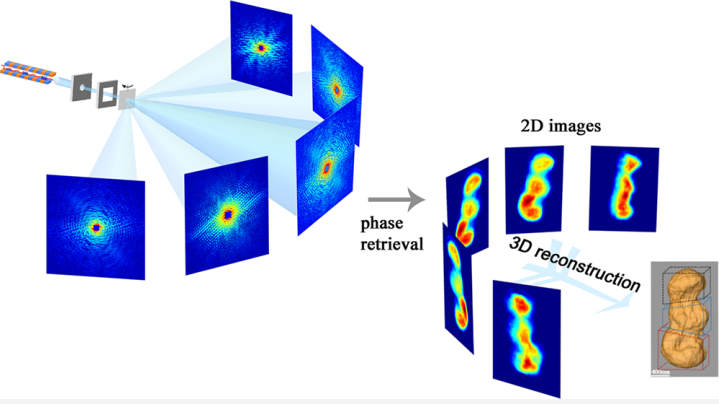
The joint team performed specific targeting performance of Au-cluster probes against S. aureus and confirmed that the Au-cluster probes has dose-dependent bactericidal activity in vitro. Then, the coherent X-ray diffraction imaging method was used to perform 3D high-resolution quantitative imaging of probe-treated S. aureus cells (3D resolution reached 47 nm), revealing the surface and internal structure of S. aureus at the nanoscale. Compared with the control S. aureus, the surface of probe-treated S. aureus has obvious depression, the surface to volume ratios is increased, the electron density is decreased, and the loss of bacterial content is very serious, resulting in complete cell collapse and folding of the cell wall. Therefore, with the advantages of the novel coherent X-ray diffraction imaging method in high-resolution quantitative bioimaging, this study was able to visually demonstrate the antibacterial mechanism of bacterial cell wall destruction and cytoplasmic components released from cells caused by Au-cluster probes.
The paper is titled Three-Dimensional Quantitative Coherent Diffraction Imaging of Staphylococcus aureus Treated with Peptide-Mineralized Au-Cluster Probes. Li Tangmeng and He Bo, 2018 graduate students of the School of Physical Science and Technology, ShanghaiTech University are the co-first authors of the paper, and professor Gao Xueyun of Beijing University of Technology, associate researcher Fan Jiadong and professor Jiang Huaidong of ShanghaiTech University are the co-corresponding authors.
Paper link: https://pubs.acs.org/doi/10.1021/acs.analchem.2c02638